| Members: | Prof. Stella W. Pang (EE) - Project Leader
Dr. David S. K. Chiu (BMS) Dr. Johnny Ho (AP) Prof. Edwin Y.B. Pun (EE) Dr. Patrick Lee (SEE) |
The localized surface plasmon resonance (LSPR) effect was used to distinguish cell concentration on ordered arrays of Au nanoparticles (NPs) on glass substrates. Human-derived retinal pigment epithelial RPE-1 cells with flatter bodies and higher confluency were compared with breast cancer MCF-7 cells. Nanosphere lithography was used to form Au NPs with average diameters of 500 and 60 nm in order to compare cell detection range, resonance peak shift, and cell concentration sensitivity. A larger cell concentration range was detected on the larger 500 nm Au NPs compared to 60 nm Au NPs (8.56×10³ – 1.09×10⁶ vs. 3.43×10⁴ – 2.73×10⁵ cells/ml). Resonance peak shift could distinguish RPE-1 from MCF-7 cells on both Au NPs. RPE-1 cells consistently displayed larger resonance peak shifts compared to MCF-7 cells until the detection became saturated at higher concentration. For both types of cells, higher concentration sensitivity in the range of ~104 – 106 cells/ml was observed on 500 nm compared to 60 nm Au NPs. Our results show that cells on Au NPs can be detected at a large range and low concentration. Optimal cell sensing can be achieved by altering the dimensions of Au NPs according to different cell characteristics and concentrations.
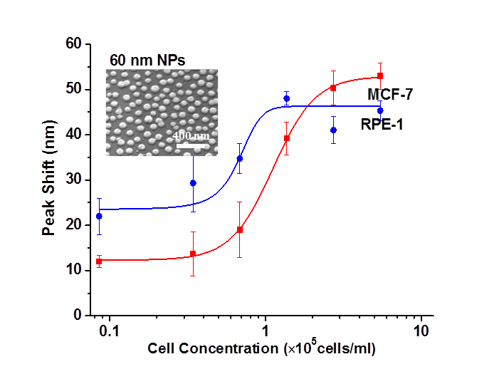
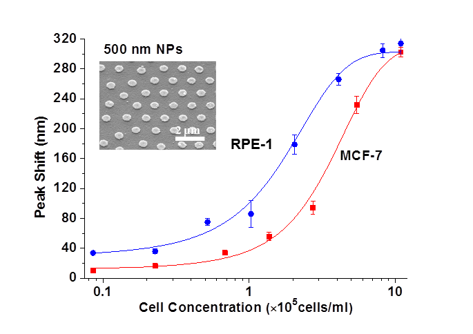
LSPR peak shift as a function of RPE-1 and MCF-7 cell concentration in the range 8.56×10³ to 1.09×10⁶ cells/ml for 60 nm (left) and 500 nm (right) Au NPs.
|
|
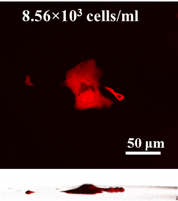
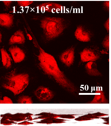
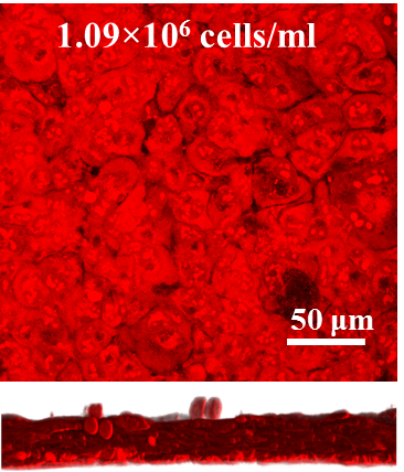
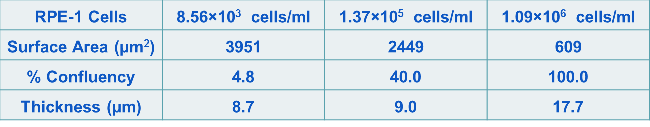
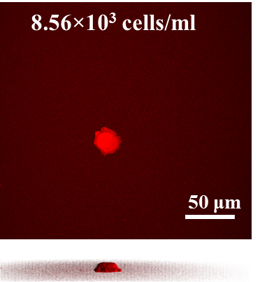
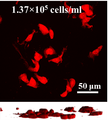
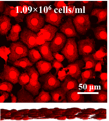
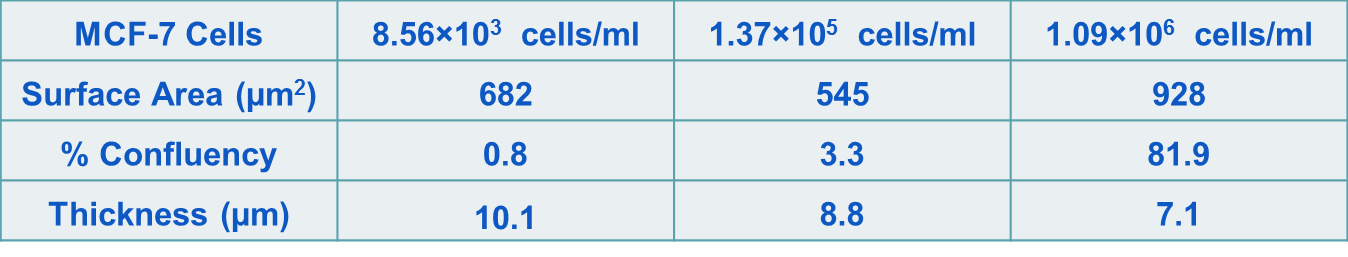
Maximum projections of 30 fluorescent confocal micrographs of the X-Y plane of RPE-1 and MCF-7 cells at different confluency seeded on 60 nm Au NP coated glass substrates with cell concentration varying from 8.56×10³ to 1.09×10⁶ cells/ml.
Publication and Conference Presentation